Welcome to the Anatomy and Physiology Lab Manual! This guide provides comprehensive resources, including answer keys and detailed exercises, to enhance your understanding of human anatomy and physiology through hands-on laboratory experiences.
1.1 Overview of the Lab Manual Structure
The Anatomy and Physiology Lab Manual is organized into chapters, each focusing on specific systems like skeletal, muscular, and nervous systems. Each chapter includes pre-lab assignments, detailed procedures, and post-lab questions. The manual also provides answer keys for exercises, ensuring self-assessment and understanding. Additional resources, such as visual guides and practice tests, are included to enhance learning and preparation for lab sessions.
1.2 Importance of Lab Work in Anatomy and Physiology
Lab work is essential for understanding anatomy and physiology, offering hands-on experience with tissues, organs, and systems. It enhances visual learning through microscopy and dissection, reinforcing theoretical concepts. Practical exercises, such as identifying tissue types and analyzing physiological processes, prepare students for real-world applications. Access to answer keys and lab manuals ensures accurate self-assessment and improves retention of complex biological principles.
Histology and Microscopy
Histology involves studying tissues under a microscope to understand their structure and function. Mastering microscopy skills is crucial for identifying cellular details and tissue types accurately.
2.1 Understanding Microscope Components and Usage
Mastering the microscope begins with identifying its components: eyepiece, objective lenses, stage, focus knobs, and condenser. Proper usage involves preparing slides, adjusting light intensity, and focusing step-by-step to observe specimens clearly.
2.2 Identifying Tissue Types Under a Microscope
Under the microscope, epithelial tissues appear as tightly packed cells in sheets or layers, while connective tissues display a loose or dense matrix with scattered cells. Muscle tissues are recognized by their elongated, striated fibers, and nervous tissues show densely packed neurons with extensions. Proper staining and magnification are essential for accurate identification. Practice with prepared slides and reference images to enhance your proficiency in tissue recognition.
Skeletal System
The skeletal system provides structural support, protects internal organs, and facilitates movement. It comprises bones and joints, with classifications based on shape and function, enabling diverse physiological roles.
3.1 Bone Structure and Classification
Bones are composed of compact and spongy bone tissue, with a periosteum covering the outer surface. They are classified into long, short, flat, irregular, and sesamoid types based on shape and function. Long bones, like the femur, support body weight, while flat bones, such as the skull, provide protection. This classification aids in understanding their structural and physiological roles in the skeletal system.
3.2 Axial and Appendicular Skeleton Analysis
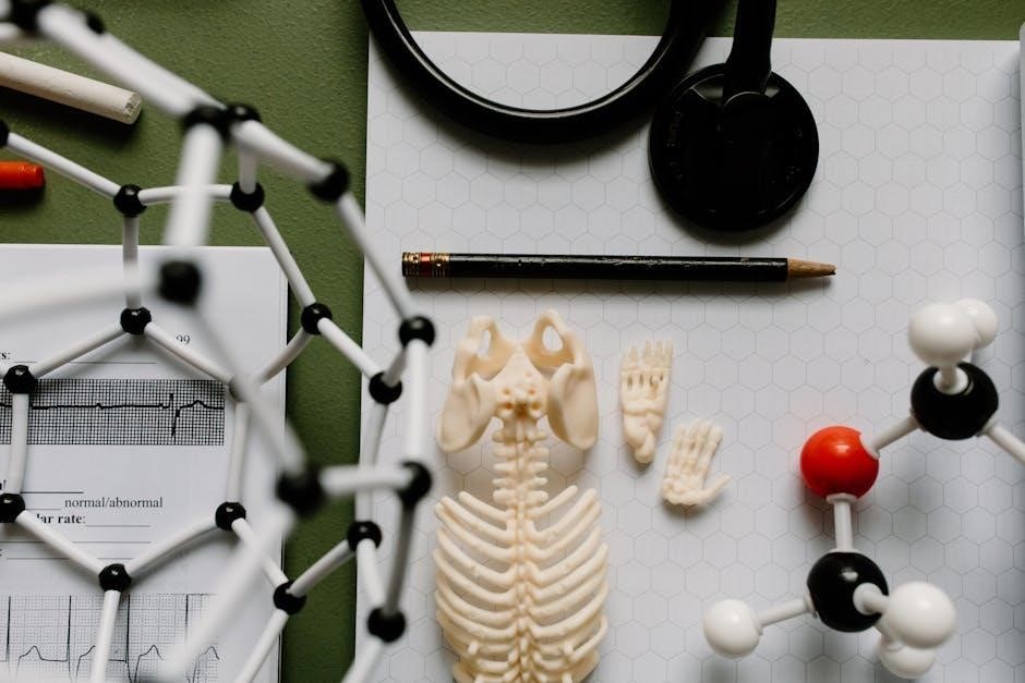
The axial skeleton includes the skull, vertebral column, ribs, and sternum, forming the body’s central framework and protecting vital organs. The appendicular skeleton comprises the upper and lower limbs, pelvis, and shoulder girdles, enabling movement and support. Analysis of these systems involves identifying bones, understanding their functions, and exploring how they contribute to overall skeletal integrity and mobility in the human body.
Muscular System
The Muscular System consists of three types of muscles: skeletal, smooth, and cardiac. It enables movement, maintains posture, and supports bodily functions like circulation and digestion.
4.1 Types of Muscles and Their Functions
The muscular system comprises three primary types of muscles: skeletal, smooth, and cardiac. Skeletal muscles are voluntary, attached to bones, and enable movement and posture. Smooth muscles are involuntary, found in internal organs, and regulate processes like digestion. Cardiac muscle is specialized for the heart, ensuring rhythmic contractions. Each type has distinct structures and functions, working together to support movement, maintain posture, and facilitate essential bodily functions like circulation and respiration.
4.2 Muscle Physiology and Movement Mechanisms
Muscle contraction occurs through the sliding filament theory, where actin and myosin filaments interact. Sarcomeres, the structural units of muscle fibers, shorten during contraction. This process is regulated by nerve impulses, triggering calcium release and muscle fiber stimulation. Movement mechanisms involve coordinated contractions and relaxations, enabling voluntary and involuntary actions. Effective muscle function relies on proper neural control and energy supply, ensuring precise and efficient movement.
Nervous System
The nervous system consists of the central and peripheral nervous systems, functioning to transmit and process information through neurons and synapses. It enables sensory perception, motor responses, and coordination of bodily functions, ensuring effective communication and control.
5.1 Structure and Function of Neurons
Neurons are specialized cells designed for communication, consisting of dendrites, a cell body, and an axon. Dendrites receive signals, while the axon transmits them. The cell body contains the nucleus and organelles. Myelin sheaths on axons enhance signal speed. Neurons communicate via synapses, releasing neurotransmitters to adjacent cells, enabling control of bodily functions, sensory perception, and information processing within the nervous system.
5.2 Sensory and Motor Responses in the Nervous System
The nervous system processes sensory and motor responses through afferent and efferent pathways. Sensory neurons transmit stimuli to the central nervous system, while motor neurons carry responses to effectors like muscles and glands. This reflex arc enables rapid reactions, such as withdrawing from pain, without conscious thought, demonstrating the nervous system’s efficiency in maintaining homeostasis and enabling voluntary actions.
Digestive and Circulatory Systems
The digestive system processes nutrients through organs like the stomach and intestines, while the circulatory system transports oxygen and nutrients via blood circulation, sustaining life.
6.1 Digestive System Organs and Processes
The digestive system consists of organs like the mouth, esophagus, stomach, small intestine, and large intestine, which break down food into nutrients. The mouth initiates digestion with teeth and enzymes, while the stomach uses acids and gastric juices. The small intestine absorbs nutrients into the bloodstream, and the large intestine manages water absorption and waste elimination. This process ensures proper nutrient uptake and waste removal, essential for overall health.
6.2 Blood Composition and Circulation Analysis
Blood is composed of plasma, red blood cells (RBCs), white blood cells (WBCs), and platelets. Plasma carries nutrients, hormones, and waste products, while RBCs transport oxygen. Circulation involves the heart pumping blood through arteries, veins, and capillaries, ensuring oxygen and nutrient delivery to tissues. Lab exercises analyze blood components and their functions, aiding in understanding circulatory mechanisms and their role in maintaining homeostasis.

Respiratory System
Welcome to the Respiratory System section! This chapter explores the essential functions of breathing, gas exchange, and oxygen transport. Lab exercises and answers guide you through analyzing lung structure, respiratory muscles, and the process of respiration, helping you master the mechanisms that sustain life.
7.1 Lung Structure and Breathing Mechanisms
The lung structure includes alveoli, bronchioles, and trachea, essential for gas exchange. Breathing mechanisms involve the diaphragm and intercostal muscles, enabling inhalation and exhalation. Lab exercises provide detailed answers to understanding how these components work together to facilitate oxygen intake and carbon dioxide removal. This section helps students visualize and analyze the respiratory process through hands-on activities and clear explanations.
7.2 Gas Exchange and Oxygen Transport
Gas exchange occurs in the alveoli, where oxygen diffuses into blood and carbon dioxide diffuses out. Oxygen binds to hemoglobin in red blood cells for transport. Lab exercises provide detailed answers on how concentration gradients and capillary networks facilitate this process. Understanding these mechanisms is crucial for grasping respiratory physiology and its role in maintaining life-sustaining functions.
Urinary and Reproductive Systems
This section explores the urinary and reproductive systems, focusing on their structures, functions, and physiological processes. Lab exercises provide detailed answers to help students master these complex systems.
8.1 Kidney Function and Urine Formation
The kidneys play a vital role in filtering blood, removing waste, and regulating fluid balance. This section explores the processes of glomerular filtration, tubular reabsorption, and secretion, essential for urine formation. The lab manual provides detailed answers to questions about nephron structure and function, along with exercises to analyze histological slides and physiological data, ensuring a comprehensive understanding of kidney physiology.
8.2 Reproductive Organs and Their Physiological Roles
This section delves into the structure and function of male and female reproductive organs, emphasizing their roles in gamete production, hormonal regulation, and fertilization. The lab manual provides detailed answers to exercises on reproductive physiology, including histological analysis of reproductive tissues and the study of processes like spermatogenesis and oogenesis. Understanding these mechanisms is crucial for grasping human reproductive health and function.
This manual provides a comprehensive understanding of anatomy and physiology through labs. Utilize the answer keys and resources. Stay engaged and practice for success effectively.
9.1 Key Takeaways from the Lab Manual
The lab manual offers a comprehensive resource for mastering anatomy and physiology. It provides hands-on experience with microscope usage, tissue identification, and system analysis. Key concepts include bone classification, muscle functions, and neural processes. Emphasizing practical applications, the manual enhances understanding of cellular structures and physiological mechanisms. Regular practice and review of answer keys ensure proficiency in lab exercises and prepare students for exams effectively.
9.2 Best Practices for Mastering Anatomy and Physiology
To excel in anatomy and physiology, prioritize consistent practice and active participation in lab exercises. Utilize answer keys and study guides to reinforce concepts. Incorporate visual aids, such as diagrams and simulations, to enhance understanding. Collaborate with peers for group study and discussions. Stay organized by reviewing notes and lab materials regularly. Apply theoretical knowledge to real-world scenarios to deepen comprehension and retention of complex physiological processes.
